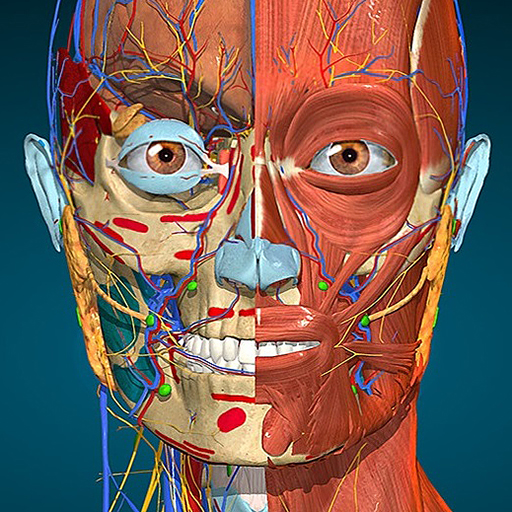
vet-Anatomy is an atlas that focuses on veterinary anatomy and is based on veterinary medical imaging.
It was created on the same platform as the renowned e-Anatomy, which is a popular medical atlas for human anatomy, especially in the radiology field. With the guidance of Dr. Susanne AEB Boroffka, ECVDI graduate, PhD, vet-Anatomy includes interactive and detailed radiological anatomy modules containing veterinary medical images, such as X-ray, CT, and MRI. The images are labeled in ten different languages, including the Latin Nomina Anatomica Veterinaria.
This application offers users various features, such as the ability to scroll through image sets by dragging their finger, zooming in and out, tapping labels to display anatomical structures, selecting anatomical labels by category, easily locating anatomical structures through the index search, and multiple screen orientations. Additionally, users can switch languages at the touch of a button.
The subscription to the application costs 89.99 € per year and includes access to all modules. It also gives users access to vet-Anatomy on the IMAIOS website. The subscription automatically renews, and users can turn off auto-renewal by going to their account settings on the Play Store after purchase. The size of the application is 750 Mb, and a Wi-Fi connection is required to download images.
There are two methods of activation for vet-Anatomy. IMAIOS members who have access to vet-Anatomy provided by their university or library can use their user account to access all modules. New users can subscribe to vet-Anatomy, and all modules and features will be active for a limited period of time.


